
Description
An Optical Coherence Tomography scan (commonly referred to as an OCT scan) is the latest advancement in imaging technology. Similar to ultrasound, this diagnostic technique employs light rather than sound waves to achieve higher resolution pictures of the structural layers of the back of the eye.
A scanning laser is used to analyze the layers of the retina and optic nerve for any signs of eye disease, similar to a CT scan of the eye. It works using light without radiation and is essential for early diagnosis of glaucoma, macular degeneration, and diabetic retinal disease.
With an OCT scan, doctors are provided with color-coded, cross-sectional images of the retina. These detailed images are revolutionizing early detection and treatment of eye conditions such as wet and dry age-related macular degeneration, glaucoma, retinal detachment, and diabetic retinopathy.
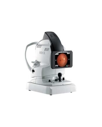
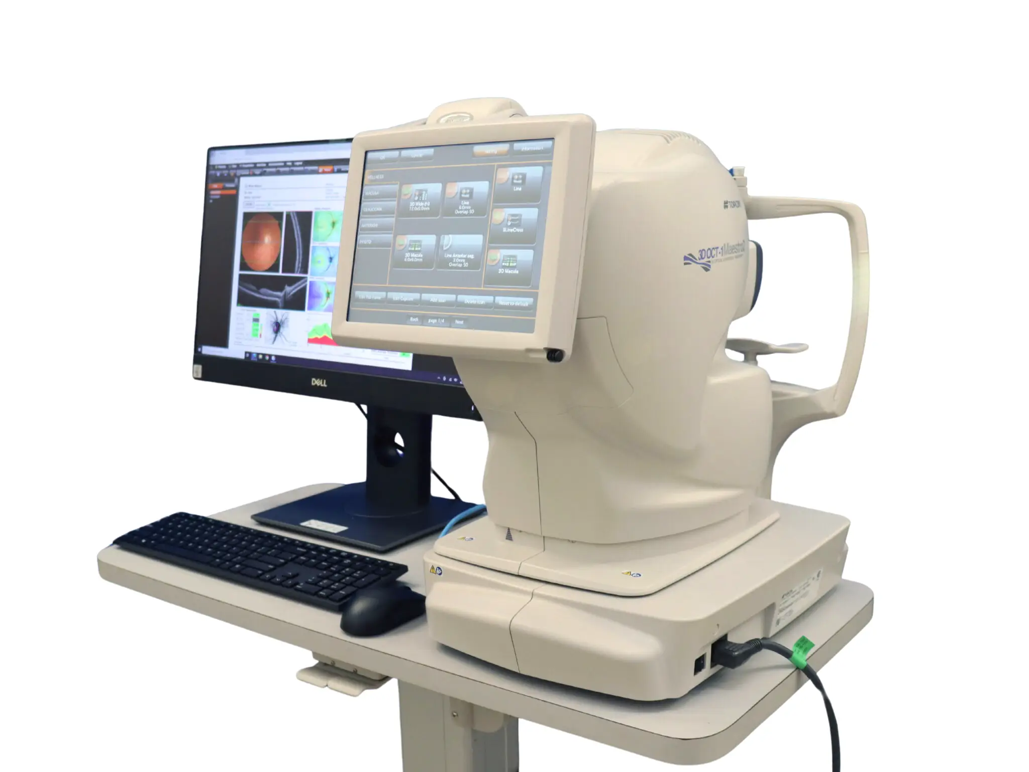
Topcon Maestro 2 OCT Fundus Camera OCTA the latest OCT technology and generates high-resolution, cross-sectional, and three-dimensional (3D) images of the retina, optic disc, and anterior segment, to give you valuable information that aids in the diagnosis and management of a range of ocular diseases.
Topcon Maestro 2 OCT scan is a non-invasive, painless test. Feel free to contact our office to inquire about an OCT at your next appointment.
Topcon Maestro 2 OCT system that automatically performs alignment, focus and capture with a single touch. The reports enables comprehensive analysis of the macula, optic disc and anterior segment. 12mm 9 mm 3D wide scan captures macula and optic disc and includes the Hood report for Glaucoma
Topcon Maestro 2 OCT Features :
– Combined OCT and color retinography
– Fully automated capture
– Compact, space-saving design
– Wide 3D scan and glaucoma module with Hood Report
– Normative database for retinal thickness, RNFL, ganglion cells and ganglion complex
– Automatic layer segmentation
– Anterior segment OCT
– Peripheral fundus photography
– 3D visualization
Topcon Maestro 2 OCT is the user-friendly OCT-Fundus Camera system that automatically performs alignment, focus and capture with a single touch. The resulting reports enable comprehensive analysis of the macula, optic disc and anterior segment. Reports can be auto exported, quickly printed or sent to your EMR in common file formats.
Automation
With the touch of a button, Maestro enables you to acquire a high resolution OCT image and a true-color fundus photo. Auto Focus, Auto Alignment, Auto Capture.
Speed of Use
Maestro requires nothing more than to touch the capture icon and Start Capture button. Alignment, focus, optimizing, and capturing are performed in an automatic procedure. After the capturing, a report can be immediately displayed by clicking on the icon.
12x9mm 3D Wide Scan & the Hood Report for Glaucoma with Reference Database – Retinal Thickness/RNFL/GCL and Optic Nerve Metrics in just one scan. Additionally, the New Hood Report for Glaucoma is available – an innovative one-page report that simplifies your structure/function decision-making.
Connectivity
Connect your Topcon Maestro 2 OCT and other diagnostic instruments, whether they are DICOM or not to review your exam data on any PC or mobile device.
Fully-Featured OCT With True Color Fundus Imaging – Not Just an OCT: Powerful, versatile and fast (50K A-Scans/sec), the OCT Fundus Camera captures beautiful and sharp B-Scans, wide-encompassing disc/macula 12×9 cubes, and informative anterior segment scans – all of which are simultaneously and easily obtained with a true color image.
User-friendly
A user-friendly OCT. The Maestro2 uses robotic technology and improves practice efficiency whilst providing optimal patientcare.
Fully Automated Capture
With a single touch, the Maestro2 automatically performs alignment, focus, optimization and capture. After image capture, the report can be immediately displayed by clicking on the icon.
Manual/Semi-Automatic Capture
In addition to automated capture, the Maestro2 offers manual/semi-auto options for difficult-to-image patients.
Maestro2