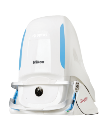
Description
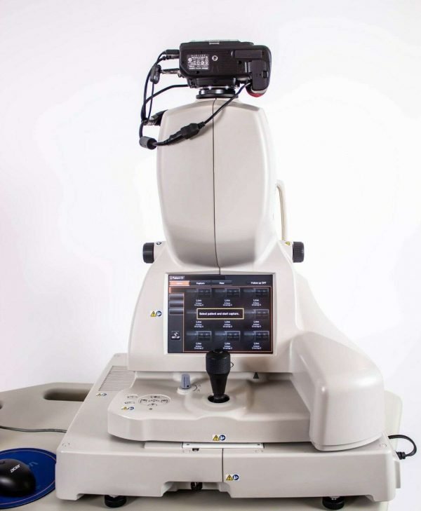
For eye care professionals who are looking for the optimal choice to help them get accurate and great quality output every time, the Topcon OCT 2000 could be an ideal model. It features high-resolution imaging to help meet the needs of practices of all sizes. The model also combines improved function with simplicity in its operations, allowing operators to develop satisfactory examination parameters with the touch of a button.
The Topcon OCT 2000 Optical Coherence Tomography features #D imaging, and presents a wide scan. It captures peripheral aspects of the macula and disc to create a more effective assessment tool.
It will improve the ability to determine the thickness and significance of different OCT images, allowing for quicker detection of macular conditions such as glaucoma. These templates are easy to read and report, with patients being better able to grasp their condition and treatment options. What features does the Topcon OCT 2000 offer?
Topcon OCT 2000 Features
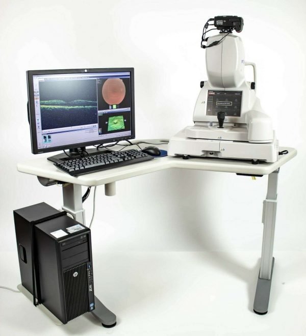
- FastMap™ software enables dynamic viewing of 2D, 3D and fundus images simultaneously
- Embedded touch-screen for quick and easy navigation
- Automated image acquisition process (auto-focus, auto-shoot)
- Data from Stratus® OCT and 3D OCT-1000 can be easily imported, analyzed and viewed
- 2 in 1 system is space-saving and economical
- Combined diagnosis with PinPoint™ registration of the OCT image within the fundus image
- Compare function allows for serial monitoring of OCT images
- Complete Windows 7 Software
- Large HD monitor
Automated function
This Topcon OCT 2000 is so automated that it practically runs itself. Your clinicians will require minimal interference during an examination or assessment because most of the functions are pretty much automated. The automatic disc searches and detect function will detect the center of the disk, which will help with the placement of the scan. Topcon OCT 2000 also features automatic fovea center detection. It can compensate for rescanning imbalance, with automatic correction of Z movement, automatic tracking of X movement and rescanning in the event of poor scanning from mistimed Y movement.
Glaucoma-specific and Macula-specific reporting
You can choose between two types of examinations to suit your patients’ needs. The wide scan angle allows you to capture an image from the optic disc to the macula, which will increase the effectiveness of any examination. Patients will be exposed to the model for shorter times, increasing the efficiency of operation and overall level of comfort. Color fundus, OCT images, cup to disc ratio and RNFL thickness maps can be generated to help determine the diagnosis. Different 3D macular reports are also available for a comprehensive assessment.
Ease of use
The Topcon OCT 2000 is relatively easy to understand and use. It can be run by clinicians with minimal understanding of OCT systems and will require only four basic steps. After registering the patient, you will be required to select a scanning pattern from the hundreds of custom options available. You will then begin the examination and consider the results afterward. The model also features a joystick controller which is motorized for high efficiency. The joystick controller allows for non-distracted adjustment, which will enable the operator to make precision movements without looking away from the screen.
High-quality output
The 50,000 A-scans per second will provide a great amount of detail in a short time. It also features a wide scan surface and higher resolution images. With different imaging variations, operators can develop a scan to suit the needs of their patients every time. This will allow for improved efficiency and performance since clinicians who are looking for a simple overview can focus on simple scans while those interested in detailed examinations can find more detailed scan options.
- Integrated, high resolution (12.3MP) fundus camera
- FastMap™ software enables dynamic viewing of 2D, 3D and fundus images simultaneously
- Embedded touch-screen for quick and easy navigation
- Historic patient data from Stratus® OCT can be easily imported, analyzed and viewed
- Seamless integration with EyeRoute® Image Management System
Topcon OCT 2000 include
- 3 years warranty
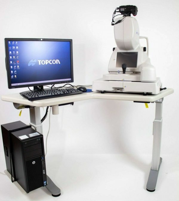
- Ergonomic power table
- Nikon D700 camera
- Large HP monitor
- Computer with the latest software

