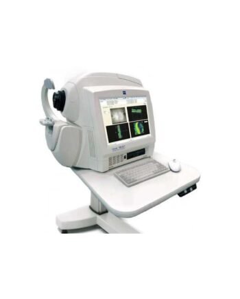

Description
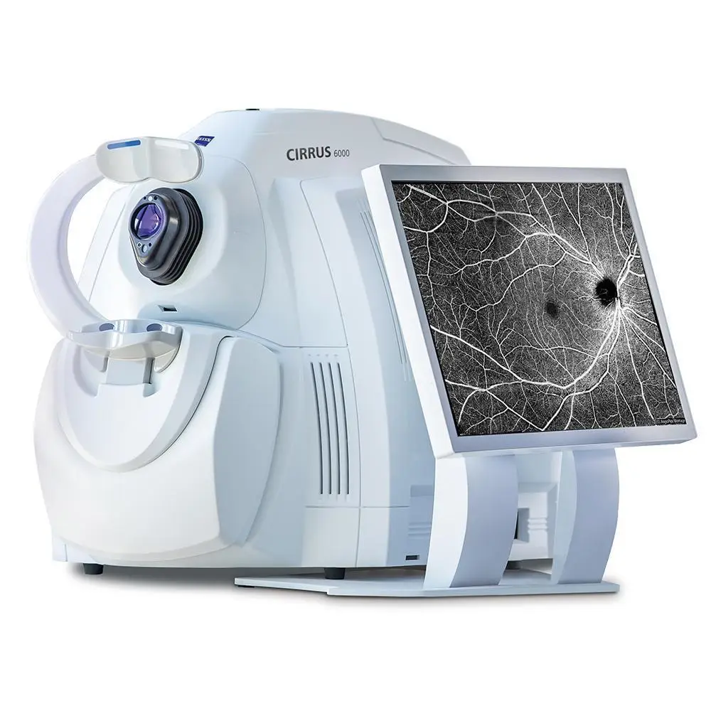
ZEISS Cirrus 6000 AngioPlex OCT Angiography ushers in a new era of eye care with non-invasive imaging of retinal microvasculature, taking diabetic eye disease management and treatment planning to the next level.
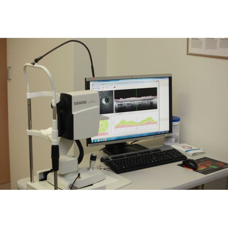
For patients with diabetic retinopathy, ZEISS Cirrus 6000 AngioPlex can help highlight early signs of diabetic changes through quantitative examination of the foveal avascular zone (FAZ) and instances of microaneurysms that may not be clearly demonstrated on conventional OCT. ZEISS Cirrus OCTA is Forum compatible, allowing the clinician the ability to overlay the OCTA data cube over the ZEISS Clarus true colour ultra-widefield image and create a full analysis in a single screen. This also provides a clear patient education visualisation to assist in the initial assessment and treatment of diabetic eye disease.
The large monitor of the Cirrus 6000 displays ultra-clear, high-definition scans to help you assess your patient’s condition and identify subtle changes in pathology. Cube data allows you to analyze data from multiple views for comprehensive insight, and Smart HD scans automatically center on the fovea, targeting critical areas. Additionally, the Cirrus 6000 introduces AngioPlex OCT angiography option to provide non-invasive 3-D microvascular imagery.
ZEISS Cirrus 6000 AngioPlex Performance
100,000 scans per second to power your practice
CIRRUS® 6000 is the next-generation OCT from ZEISS, delivering high-speed image capture with HD imaging detail and a wider field of view so you can make more informed decisions and spend more time with the patients who need it.
Faster, wider with a new level of detail
At 100,000 scans per second, ZEISS CIRRUS 6000 enables clinicians to image a larger field of view up to 12mm in a single scan. It also captures high-definition (HD) OCT revealing the finer microvascular details of the retina and providing more insight into your patient’s condition.
Making the revolutionary, routine
ZEISS Cirrus 6000 AngioPlex ushers in a new era of eye care with non-invasive imaging of retinal microvasculature – taking glaucoma and retinal disease management and treatment planning to the next level. By offering the industry’s most comprehensive tools for assessing and analyzing a range of pathologies, ZEISS provides a complete OCT Angiography (OCTA) solution.

ZEISS Cirrus 6000 AngioPlex Technical specifications
Key Parameters
Methodology: Spectral domain OCT
Optical source: Superluminescent diode (SLD), 840 nm
A-scan depth: 2.0 – 2.9 mm (in tissue)
Scan speed: 100,000 A-scans per second
Min. pupil diameter: 2.0 mm
Resolution:
• Axial resolution
• Transverse resolution
5 μm (in tissue), 1.95 μm (digital)
15 μm (in tissue)
Refractive error adjustment: -20D to +20D (dopters)
Resolution:
• Axial resolution
• Transverse resolution
5 μm (in tissue), 1.95 μm (digital)
15 μm (in tissue)
Posterior Segment scans:
• OCT
• OCTA
Cube scan (Macula and Optic Disc)
HD Raster (1, 5, 21-line, cross and radial); Raster scan length 3-12 mm;
image averaging up to 100x
3×3, 6×6, 8×8, 12×12 mm (Macula); 4.5×4.5 mm (Optic Nerve Head);
14×10 mm (Montage), 14×14 mm (Montage)